Malignant Phyllodes Tumor Cytology / Unusual Histopathology In Malignant Phyllodes Tumours Causing Diagnostic Difficulties In Cytology A Retrospective Study Semantic Scholar : Apart from these, one case of phyllodes tumor and one case of giant fibroadenoma were reported.
Malignant Phyllodes Tumor Cytology / Unusual Histopathology In Malignant Phyllodes Tumours Causing Diagnostic Difficulties In Cytology A Retrospective Study Semantic Scholar : Apart from these, one case of phyllodes tumor and one case of giant fibroadenoma were reported.. 72 patients with pathologically proven fibroadenomas and 70 patients with pathologically proven phyllodes tumor. In nine cases of histologically benign tumors and one case of malignant phylloides tumor, the findings on physical examination, mammography, sonography, and aspiration biopsy were correlated retrospectively with the histologic diagnosis of resected specimens. phyllodes tumors of the breast are rare, accounting for less than 1% of all breast tumors. In addition to stromal cells, phyllodes tumors can also contain cells from the ducts and lobules. Background phyllodes tumor (pt) of the breast is an uncommon fibroepithelial neoplasm.
They tend to grow quickly, but they rarely spread outside the breast. The luminal cell is epithelial. phyllodes tumors and fibroadenomas, which may resemble each other. Study design eight fnac cases of pt (five benign and three malignant) were reviewed. Apart from these, one case of phyllodes tumor and one case of giant fibroadenoma were reported.
Giant Malignant Phyllodes Tumor Of The Breast A Rare Case Report And Literature Review from www.spandidos-publications.com Ihc can aid in visualizing the myoepithelial layer. On fine needle aspiration cytology, a diagnosis suggestive of an atypical cytology (c3) was given and patient was advised to undergo urgent biopsy and on histopathological examination a possibility of malignant phyllodes tumor was rendered. In nine cases of histologically benign tumors and one case of malignant phylloides tumor, the findings on physical examination, mammography, sonography, and aspiration biopsy were correlated retrospectively with the histologic diagnosis of resected specimens. Phylloides tumor is a rare fibroepithelial breast tumor that occasionally has unpredictable clinical behavior. malignant phyllodes tumours of the breast show clinical and mammographic signs comparable to those of benign lesions. Ck5/6, sma, p63 and calponin. However, the median age of presentation is 45 years. To elucidate the fine needle aspiration cytology (fnac) features of phyllodes tumor (pt).
The malignant phyllodes tumor and liposarcomatous differentiation component had similar genetic mutations, such as tp53, tert, egfr, rara, rb1, and med12 mutations, all of which are common mutations in phyllodes tumors.
The tumor initially named cystosarcoma phyllodes is now generally known as phyllodes tumor.1 the phyllodes tumor is a rare breast tumor, accounting for less than 1% of all breast neoplasms.1 histologically, phyllodes tumors are classified as benign, borderline, or malignant based on characteristics of the stroma.2 they can be found as a breast lump at any age including adolescence.1 the peak. tumors expressed p53 whereas six of the seven malignant phyllodes tumors expressed this protein. Osseous differentiation within a phyllodes tumor is extremely rare. It is difficult to genralize about the behavior of phyllodes breast tumors. Moderate to marked cytologic atypia large, pleomorphic and spindle shaped cells or malignant squamous cells. phyllodes tumors (pts) of the breast are rare fibroepithelial neoplasms accompanied by overgrowth of stromal cells. Background phyllodes tumor (pt) of the breast is an uncommon fibroepithelial neoplasm. In nine cases of histologically benign tumors and one case of malignant phylloides tumor, the findings on physical examination, mammography, sonography, and aspiration biopsy were correlated retrospectively with the histologic diagnosis of resected specimens. Most phyllodes tumors are benign (not cancerous), some are malignant (cancerous), and some are borderline. The luminal cell is epithelial. Learn more about phyllodes tumors. Two months later, she developed massive left pleural effusion. phyllodes tumors are a fibroepithelial tumor composed of an epithelial and a cellular stromal component.
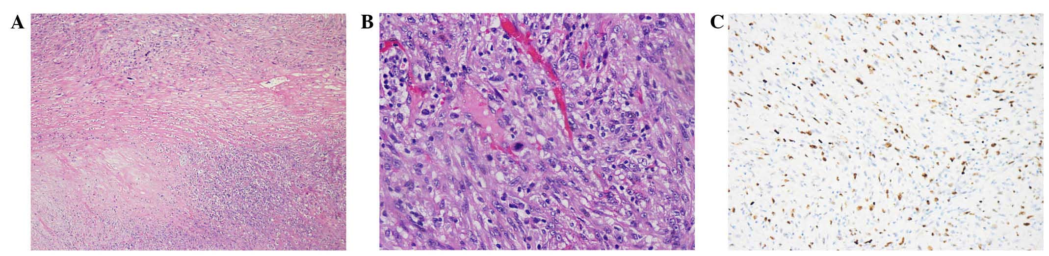
Based on these findings, our data is compatible with figure 2 microscopic findings. Mckenzie*, djumadi achmed* and c. Can phyllodes tumours of the breast be distinguished from fibroadenomas using fine needle aspiration cytology? The fine needle aspiration (fna) cytology can diagnose the phyllodes with an accuracy of only 63% which can rich up to 92.8%. These results indicate that the pathogenesis of the pleomorphic liposarcomatous differentiation was similar with that of.
Phyllodes Tumor Of The Breast With Mixed Liposarcoma Ott from www.dovepress.com Pst are classified into stromal tumor of uncertain malignant potential (stump) and pss in 2004 who classification of prostate tumors. A large tumor in the right breast was resected by simple mastectomy and diagnosed histologically as being a malignant phyllodes tumor. The diagnosis is confirmed by histology, treatment is based on surgery, which may be a large lumpectomy or mastectomy, and the prognosis depends on several factors, the most important of which is the margin for surgical resection. They may be considered benign, borderline, or malignant depending on histologic features including stromal cellularity, infiltration at the tumor's edge, and mitotic activity. The world health organization (who) has classified pts histologically as benign, borderline, and malignant. As both phyllodes tumours and fibroadenomas belong to a spectrum of fibroepithelial lesions, accurate cytological diagnosis of phyllodes tumours by fine needle aspiration can be difficult.43 44cytologically, it is often easier to differentiate benign from malignant phyllodes tumours than to separate benign phyllodes tumours from fibroadenomas.45 the presence of. phyllodes tumors and fibroadenomas, which may resemble each other. Can phyllodes tumours of the breast be distinguished from fibroadenomas using fine needle aspiration cytology?
phyllodes tumors can either be benign, suspicious of malignancy or overtly malignant.indeed, the diagnosis will depend upon the specific histological findings in.
In nine cases of histologically benign tumors and one case of malignant phylloides tumor, the findings on physical examination, mammography, sonography, and aspiration biopsy were correlated retrospectively with the histologic diagnosis of resected specimens. The luminal cell is epithelial. Pts can be detected in all ages; In addition to stromal cells, phyllodes tumors can also contain cells from the ducts and lobules. phyllodes tumors can either be benign, suspicious of malignancy or overtly malignant.indeed, the diagnosis will depend upon the specific histological findings in. At one extreme, malignant phyllodes tumours, if inadequately treated, have a propensity for rapid. tumors expressed p53 whereas six of the seven malignant phyllodes tumors expressed this protein. As with all stromal tumors, ample sampling is necessary because the sarcomatous component may be small and limited to a small portion of the tumor. They tend to grow quickly, but they rarely spread outside the breast. In an attempt to determine whether it is possible to distinguish phyllodes tumours (pts) of the breast from fibroadenomas (fas) using fine needle aspiration cytology (fnac), we reviewed the cytological slides of eight histopathologically confirmed pts (six benign and two malignant) and compared them with cytological features of 13. Were evaluated in this retrospective study. All forms of phyllodes tumors are regarded as having malignant. According to histopathological characteristics, such as tumor margins, mesenchymal cell numbers, interstitial cell atypia, mitotic activity, interstitial overgrowth, and malignant heterogeneous elements, the world health organization classifies pts into benign.
Cytological and histological findings of a case of malignant phyllodes tumor with osseous differentiation are presented. Ck5/6, sma, p63 and calponin. Moderate to marked cytologic atypia large, pleomorphic and spindle shaped cells or malignant squamous cells. World health organization divided phyllodes tumor into benign, borderline, and malignant categories based on the degree of stromal cellular atypia, mitotic activity per high power elds, degree of stromal phyllodes tumors are a fibroepithelial tumor composed of an epithelial and a cellular stromal component.
Case 08 from www.bccancer.bc.ca Sarcomatous differentiation in a malignant phyllodes tumor: Most phyllodes tumors are benign (not cancerous), some are malignant (cancerous), and some are borderline. Mckenzie*, djumadi achmed* and c. The patient developed local recurrence 4 months later while on adjuvant radiotherapy and she had a salvage resection. phyllodes tumors are a fibroepithelial tumor composed of an epithelial and a cellular stromal component. The diagnosis is confirmed by histology, treatment is based on surgery, which may be a large lumpectomy or mastectomy, and the prognosis depends on several factors, the most important of which is the margin for surgical resection. Ihc can aid in visualizing the myoepithelial layer. The histologic diagnosis was malignant phyllodes tumor (pt).
Osseous differentiation within a phyllodes tumor is extremely rare.
The histologic diagnosis was malignant phyllodes tumor (pt). phyllodes tumors are more likely to become malignant and should be evaluated for mitotic rate and histological characteristics. The malignant phyllodes tumor and liposarcomatous differentiation component had similar genetic mutations, such as tp53, tert, egfr, rara, rb1, and med12 mutations, all of which are common mutations in phyllodes tumors. Following this, lumpectomy was performed and a diagnosis of malignant phyllodes tumor was confirmed. A phyllodes tumor, which derives from the greek word 'phullon' The diagnosis is confirmed by histology, treatment is based on surgery, which may be a large lumpectomy or mastectomy, and the prognosis depends on several factors, the most important of which is the margin for surgical resection. Cytological and histological findings of a rare case of a malignant phyllodes tumor (with liposarcomatous components) of the breast are presented. Preoperative diagnosis with fine needle aspiration cytology. A large tumor in the right breast was resected by simple mastectomy and diagnosed histologically as being a malignant phyllodes tumor. In contrast, benign phyllodes tumors on clinical, radiological, and cytological examination are often indistinguishable from fibroadenomas and can be cured by local surgery. Single or clustered spindled cells with nuclear pleomorphism. Mckenzie*, djumadi achmed* and c. These results indicate that the pathogenesis of the pleomorphic liposarcomatous differentiation was similar with that of.
The world health organization (who) has classified pts histologically as benign, borderline, and malignant phyllodes tumor malignant. A large tumor in the right breast was resected by simple mastectomy and diagnosed histologically as being a malignant phyllodes tumor.


0 Comments